Neurologia e Neurocirurgia Anatomia, Biologia, Bioquímica, Embriologia, Fisiologia, Histologia e Neuroanatomia
Neuroanatomy of the Dog – Third Edition
De: Vicente Aige Gil
ISBN: 9798900500225
2026, Editoras Diversas
Capa dura
Páginas:
Neurologia e Neurocirurgia Anatomia, Biologia, Bioquímica, Embriologia, Fisiologia, Histologia e Neuroanatomia
De: Vicente Aige Gil
ISBN: 9798900500225
2026, Editoras Diversas
Capa dura
Páginas:
This second edition is an improved revision of the first edition in which two new chapters have been added (Chapter 16 is an atlas of transverse sections of the dog’s head, and Chapter 17 is an MRI atlas of the axial muscles and the vertebral column). I have also added new videos, images and few modifications in the text. The app was updated. Some of them were the result of new dissections.
This third edition is an improved revision of the second edition in which new images have been added as a result of new dissections, and some concepts revised.
The book has been divided into 18 chapters beginning with general principles in which the need to have a nervous system and its function is analyzed. It is followed by a chapter on embryological development that ranges from neurulation to malformations. The parts of the nervous system have been divided into 11 chapters that include numerous images and videos. This is followed by a chapter on the biophysical principles of MR imaging and a chapter that includes an atlas of MRI and two videos of cross-sections of the brain. It ends with a chapter intended for students on how to localize neurological lesions with some cases and 11 videos to check your knowledge.
The book covers the neuroanatomy aspects of the dog's nervous system, with specific references to the cat, that a neurologist should have. To carry it out, many macroscopic dissections have been made and, with the use of the magnifying glass for small areas never shown before. The text is completed with histological images for a better understanding of the function and with MR images. In the white matter chapter of the cerebrum, nerve fiber dissections have been performed following a successive freeze and thaw process. For a better three-dimensional understanding of the dissections, numerous videos are included accessible by QR. They are arranged at the end of the different chapters.
As in previous editions, the book is aimed at clinicians, neurology residents, and students with the clear intention of sharing the author's knowledge of neuroanatomy applied to veterinary neurology in dogs and cats. Hands on in cadaver dissection is the cornerstone to understand and learn the basic principles of the anatomy of the nervous system and its implication in function and disfunction in the whole body. I encourage the clinicians and students to approach this field by making their own dissections on cadavers before attempting the surgery.
1- General principles
2- Development
3- Cerebrum
4- Cerebellum
5- Brain stem
6- Brain blood vessels
7- Choroid plexuses and CSF
8- Spinal cord morphology
9- Spinal cord meninges
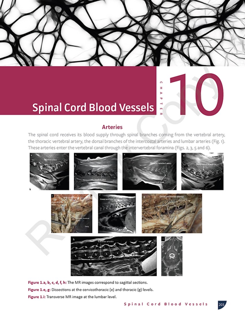
10- Spinal cord blood vessels
11- Cranial nerves
12- Spinal nerves
13- Autonomic nervous system
14- Basic Physiscs of MRI
15- MRI of the dog's head
16-Transverse head sections
17-MRI of the axial muscles
18- The neurological examination
Vicente Aige Gil, DVM, PhD
Associate Professor of Anatomy Facultad de Veterinaria. Universidad Autónoma de Barcelona. Barcelona. Spain
Graduated in Veterinary Medicine (1985), he was appointed a tenure-track position teaching anatomy at the Universidad de Zaragoza (Spain) and obtained his PhD (1988), with cum laude, with a thesis on the pineal gland of the chick embryo. After a post-doc research at the Universities of Reading (UK) and Guelph (Canada), and being a visiting professor at the Veterinary School of Glasgow (Scotland), he was appointed as senior lecturer / associate professor of veterinary anatomy at the Veterinary Faculty of the Universidad Autónoma de Barcelona (UAB). He is a member of the American Association of Veterinary Anatomists (AAVA) and an invited neuroanatomy lecturer in courses and seminars national and international.